Leaving Cert Biology Revision: Musculoskeletal System
- Javeria Saleem
- Jun 24, 2024
- 7 min read
Musculoskeletal System
The Skeleton,Muscles and Movement
Made of bone and cartilage, the human skeleton is known as the endoskeleton or internal skeleton.

Principal Roles
1. The human body's shape is primarily due to its support for the soft tissues.
2. Movement: When muscles contract, the sturdy, inflexible bones act as levers.
3. Protection: fragile critical organs are shielded by the strong bones.
3. The cranium shields the brain.
3b. The rib cage shields the lungs and heart.
3c. The backbone shields the spinal cord.
Organization
1. Axial Skeleton: consists of the ribs, sternum, backbone, and skull.
2. Appendicular Skeleton: limbs, pelvic girdle, and pectoral girdle.
Axial Skeleton
1. Skull: composed of 22 fused bones except for the mandible (lower jaw).
2. The vertebral column, also referred to as the spine or backbone. It is made up of 33 tiny bones arranged in a line: the sacrum (5), lumbar (5), thoracic (12), and coccyx (4). The sacrum and coccyx vertebrae are combined into one unit.
The flexibility of the backbone is attributed to the minor movement of the vertebrae in the other locations. Shock-absorbing cartilage discs are located in between these. They aid in the vertebrae's protection. Ligaments support the vertebrae and keep them in place.
The vertebrae are supported by muscles that are linked to their surfaces. From the spinal cord situated in between the vertebrae, spinal nerves arise in pairs.
2. Appendicular Skeleton: limbs, pelvic girdle, and pectoral girdle.

Rib Cage
Twelve pairs of ribs plus the sternum make up the rib cage. The backbone is connected to all 12 pairs. The sternum is joined to the first seven actual ribs.
The false ribs, which connect to the seventh rib, are the next three. The floating ribs are the final two. They are connected to the backbone just at one end.
The thin, flat bone in the middle of the chest wall is called the sternum or breastbone.

The Appendicular Skeleton
Two clavicles, or collar bones, and two scapulae, or shoulder blades, make up the four bones that make up the pectoral girdle. There is a ball and socket joint where the arms and scapulae meet.

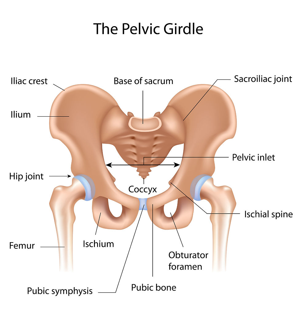
The Pelvic Girdle
The Pelvic Girdle is six fused bones that give the appearance of being one huge cylindrical bone. At a ball and socket joint, the legs and pelvis articulate. The sacrum is where the pelvis and backbone are firmly joined.

Limbs
Arms and legs have comparable construction. Long bones make up the upper skeleton. It is the arm's humerus and the leg's femur. The other two long bones in the medial region are the tibia and fibula in the leg and the ulna and radius in the arm.
The hand and foot share comparable bones. The carpals are found in the wrist of the arm, and the tarsals are found in the ankle of the leg. The metacarpals are found in the hand's palm, and the metatarsals are found in the rear foot. The phalanges are found in the fingers and toes.

Long Bone Structure
Long bones are enclosed by a membrane known as the periosteum. It is made up of nerves and blood vessels.
The shaft, or long major part, of the bone is called the diaphysis. The diaphysis forms after the epiphysis, which is located at each end of the long bone.
On either end of the long bones lies cartilage. Collagen is the protein that makes it up. The surrounding material of calcium and phosphorus salts is coiled around collagen fibers.
The epiphyses at freely moving joints are shielded from shock and friction by the cartilage covering them. There are no blood vessels or nerves in the cartilage.
Diffusion allows beneficial substances to enter the cartilage.
Bone Types
Compact and spongy bones are the two different forms of bones.
A solid bone with a concentric ring structure at the microscopic level. Osteoblasts are the bone cells that make it up. These cells are developing inside a substance known as bone matrix. Thirty percent collagen and seventy percent inorganic salts, including calcium phosphate, make up the bone matrix. The protein gives the bone flexibility, while the calcium salts give it strength. The bone contains both nerve fibers and blood arteries.
2. Spongy Bone
an uneven, thin-plate assemblage of bone. Another name for it is cancellous or trabecular bone. The mineral deposits are stacked like struts in a system. In between the plates, bone marrow fills the gaps. The area of the diaphysis where marrow is also found is called the marrow cavity.

Bone Marrow
Bone marrow comes in two varieties:
The production of red blood cells, white blood cells, and platelets occurs in the red bone marrow.
The yellow bone marrow is in charge of storing energy as fat, or lipid. If the body requires more red blood cell production, this can be transformed into red marrow. Is it restricted to adults only? Medullary cavity.
Growth of Bones
Up to the eighth week of development, the embryo is composed of cartilage. After that, bone takes its place. Collagen is produced by osteoblasts.
Around the collagen is a hard layer that is primarily composed of calcium phosphate. The osteoblasts become inactive bone cells when they are trapped inside the calcium phosphate.
Growth plates between the diaphysis and the epiphysis cause the bone to elongate. Cartilage is continuously forming at this plate, eventually transforming into bone.
We refer to this process as ossification. When a person enters adulthood, this comes to an end.

Bone Formation
Although bone resorption from increased muscle activity or weight can cause a bone's thickness or width to expand throughout life, bone growth in length ceases in early adulthood. We refer to this increase in diameter as appositional growth.
Around the outside bone surface, compact bone is formed by osteoblasts in the periosteum. Osteoclasts in the medullary cavity also break down bone around the medullary cavity on its internal bone surface at the same time.
Together, these two mechanisms broaden the bone's diameter while preventing it from growing too heavy or cumbersome.
Factors influencing the growth of bone
One cause of bone stress is physical exercise. It stimulates osteoblasts. The bone grows more.
A lack of stress results in thin bones.
Bone size is increased by sex hormones and growth hormones.
Joints
A joint is where two or more bones come together. Three main categories of joints exist:
1. Fused Joints:
The coccyx, sacrum, pelvis, and skull are examples of these joints. These joints are, as their name implies, the places where joints grow or fuse. The suture is the area where they unite during growth. These joints offer defense, stability, and strength.
2. Partially Movable Joints:
These joints are situated in the space between the upper spine's vertebrae. The joints are made of cartilage. They support and shield the bones. Ligaments hold the bones together. The ligaments are firmly attached to the bones, limiting their range of motion. The spinal cord is shielded by this.
3. Joints that are Freely Moveable or Synovial:
In these joints, a hollow divides the bones, and the ends of the bones are covered in cartilage. Ligaments bind the bones together and prevent excessive bone movement.
The sleeve-like capsule connects the two bones in addition to the ligaments. The synovial cavity is enclosed by the capsule. Ligaments make up the capsule's external layer.
As was previously mentioned, ligaments regulate a range of motion and hold bones together, avoiding dislocation. The synovial membrane is the capsule's inner layer.
The lubricating synovial fluid is secreted by the synovial membrane. To stop frictional wear and tear, lubrication is necessary.

Synovial Joint Classes
1. Gliding: These joints' bones glide over one another, back and forth, and side to side. The tarsals of the ankle and the carpals of the wrist are two examples.
2. Pivot: Turning motion is possible with these joints. Examples include tilting the head between the first and second vertebrae and turning the hand's palm up or down between the ulna and lower arm's radius.
3. Hinge: These joints permit flexion and extension in a single plane. As their name suggests, they function similarly to a door's hinge. Bending the elbow or knee are two examples.
4. Ball and Socket: This kind of joint allows for motion in all three planes, or directions. Shoulder and hip joints are two examples.

Linkages
At joints, strong, slightly elastic tissues called ligaments hold one bone to another. Warming up makes these tissues more pliable. For this reason, you should warm up gradually before working out. Ligaments regulate how far the bones may travel at a joint and stop it from dislocating.
tendon
Strong, inelastic bands of connective tissue called tendons connect muscle to bone. They are made of collagen and have blood arteries in them. Muscle contraction will not cause the inelastic tendon to stretch. Consequently, the entire pull is transferred to the bone, allowing for the completion of the entire range of motion.
Arthritic
Rheumatoid arthritis and osteoarthritis are the two kinds of arthritis. The swelling and inflammation of the joint are present in both of these disorders. Refer to page 352 of your textbook for additional details on this subject.
musculature
Muscle comes in three varieties: cardiac, smooth, and skeletal.
Skeletal muscle
As the name suggests, skeletal muscle is a muscle that is joined to the skeleton. Another name for it is striated muscle. Skeletal muscle contractions are controlled voluntarily. The body moves mostly because of these muscles. Maintaining posture, supporting the joints, and producing heat are other goals. Skeletal muscle may contract quickly and forcefully, but it also becomes tired rapidly.
Sleek Muscle
All of the body's hollow organs, except the heart, have walls made of smooth muscle. These structures get smaller as a result of their shrinkage. As a result, it controls blood flow in the arteries, the passage of food through the digestive system, the release of urine from the bladder, the expulsion of infants from the uterus, and the regulation of airflow through the lungs. Smooth muscle contractions are not controlled voluntarily. We refer to it as involuntary muscle. It tires slowly and contracts gently.
Heart Muscle
Heart muscle makes up your heart. There is only one kind of muscle in your body: the heart. Cardiac muscle is different from other muscle groups in that it never tires. It never stops operating automatically or for a break. Your heart's cardiac muscle contracts to pump blood out of it and relaxes to pump blood back into it.
Opposing Skeletal Muscle Groups
Muscle pairs make up antagonistic muscles. One member's behavior is in opposition to the other's. Although muscles can shorten (stretch), they are unable to extend themselves. They are paired so that a skeleton can transfer the contraction of one muscle or muscle group to the stretching of another muscle or muscle group. Antagonistic muscle pairs are those that stretch one another.
An antagonistic muscle pair
The arm's triceps and biceps muscles are an illustration of an antagonistic pair. The arm is brought closer to the body when the biceps are contracted, and the triceps are stretched. The arm and biceps are extended when the triceps contract.


Comments